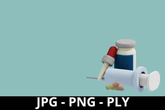
Evaluating 3D Rendering Medical Background: What You Need to Know Before Choosing
When you’re comparing visual tools for medical communication, the term 3D rendering medical background often surfaces. It refers to computer-generated three-dimensional imagery derived from patient scan data (like CT or MRI) or created from scratch to represent anatomical structures. Unlike a standard photograph or a two-dimensional medical illustration, a 3D rendering medical background offers a rotatable, often interactive model that can be segmented, labeled, and manipulated. For professionals and patients alike, it serves as a bridge between raw data and intuitive understanding. But as with any tool, it comes with specific tradeoffs that may or may not align with your project, budget, or audience.
How 3D Rendering Medical Background Differs from Conventional Medical Imaging
Standard medical images—X-rays, ultrasound sweeps, or even stacks of MRI slices—provide essential diagnostic information, but they require a trained eye to interpret spatial relationships. A 3D rendering medical background takes that same volumetric data and reconstructs it into a coherent, three-dimensional form. The difference is immediate: depth, rotation, and the ability to isolate individual tissues by adjusting transparency. For example, a radiologist viewing a series of axial slices must mentally align each layer; a 3D rendering presents the whole structure as a single object that can be viewed from any angle. This shift from abstract slices to a tangible model is what makes the 3D rendering medical background so valuable for surgical planning and patient education. However, it also introduces new considerations: the rendering process requires specialized software, adequate computational power, and someone skilled in segmentation and cleanup. Without these resources, a raw DICOM dataset might be more practical than a polished 3D rendering.
Strengths: When Detail and Interactivity Matter
The primary advantage of a 3D rendering medical background is its ability to convey complex anatomy in an intuitive way. Surgeons can rotate a model of a fractured pelvis to plan screw placement before entering the OR. Educators can highlight the course of a cranial nerve through a translucent skull. Patients who struggle with flat diagrams often grasp their diagnosis faster when they see a 3D rendering medical background that matches their own scan. Additionally, because the rendering is digital, it can be shared instantly across devices, embedded in apps, or presented in virtual reality—no physical models to store or ship. For research presentations, a well-done 3D rendering medical background adds credibility by showing spatial accuracy rather than stylized art.
Limitations: The Hidden Costs and Caveats
Despite its benefits, a 3D rendering medical background is not a universal solution. Creating a high-quality model demands time. Segmenting each organ, bone, or vessel from a scan can take hours even with automatic tools. The software itself may require expensive licensing or subscription fees. Furthermore, the rendering is only as good as the input data: poor scan quality, motion artifacts, or limited resolution will degrade the final model. There is also the risk of over-interpretation—a rendered model may appear more definitive than the underlying data actually is. For instance, a smooth surface on a 3D rendering medical background might imply a clear tumor margin, while the actual histology could be more diffuse. Medical professionals must treat these renderings as visual aids, not definitive representations. In settings where speed is critical—such as an emergency room—waiting for a full rendering may not be feasible compared to looking at the raw CT slices.
Key Factors to Consider When Evaluating 3D Rendering Medical Background Options
Before choosing a 3D rendering medical background approach, assess your specific needs. Accuracy requirements come first: for surgical navigation, you need a model that aligns precisely with the patient’s anatomy, often verified by a medical physicist. For patient education, a slightly simplified rendering may be more understandable. Interactivity level is another factor: do you need a static image for a brochure, a rotatable embedded viewer for a website, or a fully immersive VR experience? Each tier requires different development effort. Budget plays a role: outsourcing a single high-quality 3D rendering medical background can cost hundreds of dollars, while building in-house capability involves software licenses and training. Expertise matters—if your team lacks familiarity with segmentation tools, you may end up with a model that misses important details. Finally, consider the audience: a layperson benefits from a clean, color-coded 3D rendering medical background, while a specialist might need one with precise measurements and labels. Weighing these factors will help you decide whether a 3D rendering fits your project or whether an alternative like a physical model, a 2D illustration, or a video overlay might serve better.
Practical Examples of 3D Rendering Medical Background in Action
Example 1 – Preoperative planning for spinal deformity: An orthopedic surgeon uses a 3D rendering medical background generated from a patient’s CT scan to simulate screw placement. By rotating the model and adjusting the view, the surgeon identifies the safest trajectory and reduces intraoperative guesswork. The rendering also helps explain the procedure to the patient, who can see exactly where the hardware will go.
Example 2 – Medical illustration for a textbook: A publisher commissions a 3D rendering medical background of the heart to show coronary artery variations. The artist creates a semi-transparent myocardium overlaid with arterial paths. This approach allows the reader to see deep structures without the clutter of multiple overlays. The final image is more accurate than a hand-drawn illustration, yet stylized enough to highlight educational points.
Example 3 – Patient education in a clinic: A rheumatologist uses a tablet to show a rheumatoid arthritis patient a 3D rendering medical background of a healthy joint compared to their affected one. The patient can pinch and zoom, seeing pannus formation and joint space narrowing. This interactive experience leads to better understanding of treatment options and medication adherence.
Making an Informed Choice: Balancing Tradeoffs
There is no single answer when deciding whether a 3D rendering medical background is the right choice. It excels where spatial understanding, detail, and interactivity are critical—surgical planning, complex anatomy teaching, and patient communication. But it introduces cost, time, and expertise barriers that simpler methods do not. For a quick reference guide or a low-budget handout, a well-labeled 2D illustration might be more practical. For hands-on learning, a 3D-printed model provides tactile feedback that a screen cannot replicate. The best approach is to match the medium to the task: use a 3D rendering medical background when you need depth and dynamic exploration, and reserve other tools when speed, simplicity, or physical presence is paramount. By considering accuracy needs, audience, budget, and skill level, you can choose a visualization strategy that genuinely supports your goals rather than following a trend. Ultimately, the most effective medical communication uses the right tool at the right moment—and the 3D rendering medical background is one powerful option in that toolkit.